Conventional radiography is traditionally used to complementforensic autopsy, serving primarily to document metallic bullet fragments, foreign bodies, fractures, and injury patterns. It is also used to aid in the determination of identity when conventional methods of identification such as fingerprinting or DNA analysis are not available or cannot be utilized.1 Radiography Conventional radiography is the most widely used postmortem radiology technique. It is used most often to locate bullet fragments and to find projectiles and foreign bodies. In child abuse and anthropologic cases, radiography is the imaging modality of choice to evaluate subtle bone detail. C-arm fluoroscopy C-arm fluoroscopy may be used to facilitate the localization and recovery of metallic fragments or foreign bodies at autopsy when they are not readily retrievable at dissection based upon the localization of the object on radiographs. It may also be used for limited angiographic assessment of vascular integrity by directly injecting the vessel of concern with iodinated contrast material under fluoroscopic observation. Radiation safety and protection measures should be strictly followed for all personnel in the vicinity of an operating C-arm unit. Multidetector computed tomography (MDCT) scanning MDCT scanning, also known as multislice computed tomography (MSCT) scanning, is emerging as the foremost cross-sectional imaging modality in forensic medicine. Its speed, ease of use, and compatibility with metallic fragments make it an excellent complement to autopsy. The cost and availability of MDCT scanners and personnel is the most important limitation in integrating cross-sectional imaging with autopsy. As an alternative to an on-site scanner, forensic facilities may choose to collaborate with local radiology practices or hospitals in order to obtain MDCT studies on specific cases. MDCT scanners use a 2-dimensional (2-D) array of detector elements that improve resolution and scan time when compared to older CT scanners. The alignment of the detectors along the long (z-axis) of the body enables the scanners to obtain 4, 8, 16, 64, 128, or more slices with each rotation of the x-ray tube. Protocols that specify the technical and anatomic parameters for obtaining scan data, reconstructing scan data into images, and reformatting images into anatomic planes may be organized for specific anatomic regions of the body similar to clinical scanning protocols, or they can be more generalized to obtain full-body data. Angiography and MDCT angiography A variety of postmortem angiography techniques have been reported to assess vascular injury and disease.2,3 The contrast agent, delivery mechanism, and injection technique can be altered based upon the location of the suspected abnormality and radiologic technique used to image during contrast injection. Angiography may be performed with radiography, C-arm fluoroscopy, or MDCT scanning. Magnetic resonance imaging (MRI) Postmortem MRI has been used to assess soft-tissue and visceral hemorrhage, ischemia, and tumors.4,5,6Although the technical complexity, expense, and availability of MRI make it more complicated to use as a routine imaging modality compared to MDCT, it provides superior contrast resolution relative to MDCT, making it a more optimal imaging modality to visualize soft tissues. Caution should be taken when imaging bodies that contain metal, because ferromagnetic substances may pose potential harm or cause significant image degradation when placed in the magnetic field of the scanner. Blunt trauma Blunt force injury is the most common form of lethal and nonlethal trauma. Postmortem MDCT scanning is useful to visualize and reconstruct blunt injury patterns before autopsy and the 3-dimensional MDCT display of head, spine, and pelvic injuries may facilitate the understanding of the mechanism of injury.7 Radiography or MDCT scanning in cases of blunt chest trauma is useful before autopsy to show pneumothorax,tension pneumothorax, and pneumomediastinum, which may go undetected during routine dissection. Pulmonary contusions are characterized by consolidation and opacification in a nonsegmental distribution, associated with the site of impact. Consolidation in the contralateral portion of the chest is indicative of a contrecoup contusion. Gunshot wounds Gunshot wounds are exquisitely depicted on postmortem MDCT images. Gunshot wound tracks are typically linear tissue defects containing gas and metallic fragments (see the 3 images below).8 Natural deaths Postmortem MDCT scanning is the most appropriate initial cross-sectional imaging technique in suspected natural deaths, because it provides a rapid anatomic survey of the head and body. Postmortem MDCT scans provide supportive information and excludes occult trauma when atherosclerotic coronary artery disease is the cause of death. The most common postmortem MDCT findings in death from myocardial infarction from atherosclerotic coronary artery disease are coronary artery calcification and pulmonary edema. The degree of luminal narrowing or the presence of arterial occlusion can only be assessed when contrast material is injected into the arterial system during MDCT imaging. MRI may be used to assess the myocardium.6,9 Deaths from aortic aneurysm rupture cause massive hemorrhage that may surround the aorta and/or extend into the mediastinum, pericardium, or pleural spaces. In this setting, high attenuation hemorrhage will be present on MDCT images. Aneurysmal dilatation of the aorta may or may not be apparent on MDCT scans, because if residual intravascular blood volume is low, the aorta may be collapsed on postmortem imaging. The postmortem MDCT findings in aortic dissection are deformity of the aortic contour, intramural hematoma, hemopericardium, and pulmonary edema. Hemopericardium, characterized by a hyperdense inner ring and hypodense outer ring is the most common MDCT finding in deaths from aortic dissection.10 Angiography is required to confidently identify the intimal flap and false lumen of the dissection when establishing the diagnosis by imaging alone. In death from intracranial hemorrhage, acute blood is seen as high attenuation (80 to 90 Hounsfield units [HU]) on MDCT scans. If the decedent survives beyond the acute stage of hemorrhage, the hemorrhage will have lower attenuation on MDCT images. Intraparenchymal hemorrhage is surrounded by vasogenic cerebral edema, which reaches its maximum at 4 to 5 days. The margins of intraparenchymal hemorrhage become less distinct over time. Burns Postmortem MDCT scanning is useful in severely burned and charred bodies that are difficult to examine. It may help identify antemortem traumatic injury and aid in localizing tissue suitable for DNA analysis.11 Partial-thickness burns may produce no significant changes in the dermis or mild irregularity of the dermis on MDCT images (see the following 2 images). Sharp force injury Conventional radiography is considered an important component in the forensic assessment of sharp force injury to help identify and aid recovery of broken knife blades and to help differentiate stab wounds from ballistic wounds.13 If knife blade fragments are present on radiography, the location of the fragment should correlate with the expected location of the wound path based upon the skin entry site. On MDCT images, the wound track may be visualized and lead to the metallic fragment. Drowning MDCT scanning closely parallels autopsy for the depiction of the anatomic findings that are supportive for the diagnosis of drowning. Sinus fluid, mastoid fluid, subglottic tracheal and bronchial fluid, and pulmonary ground glass opacity are consistently present on MDCT images.15 Sinuses may be completely filled with fluid, contain air fluid levels, or contain high attenuation sand, which layers dependently in the sinus. Fluid and/or sand may be present within the trachea and bronchi, as shown in the following 2 images. Radiology is an integral part of forensic autopsy. Radiography alone is used in most centers. However, technologic advances in cross-sectional imaging have made it possible for MDCT scanning to be used routinely with forensic autopsy. Cross-sectional imaging makes the radiologic contribution to forensic autopsy more effective and may increase both the speed and accuracy of forensic investigation.Postmortem Radiology and Imaging
Introduction
The addition of cross-sectional imaging to forensic autopsy allows the radiologist and forensic pathologist to view postmortem anatomy in 2 and 3 dimensions without dissection. Multidetector computed tomography (MDCT) scanning and magnetic resonance imaging (MRI) can be used to focus the autopsy on specific abnormalities, view injury patterns in 3 dimensions, detect occult disease or injury without dissection, and evaluate anatomic areas that are difficult to dissect.
In certain causes of death and forensic scenarios, cross-sectional imaging may be used to help the forensic pathologists decide which decedents should have an autopsy or to determine whether the autopsy should be limited or complete. In those cases that do not undergo autopsy, cross-sectional imaging findings add anatomic information to the external examination, toxicology, and biochemical findings that may have been previously used alone to determine the cause of death.
The purpose of this chapter is to discuss postmortem imaging techniques and the benefits and limitations of postmortem radiography and cross-sectional imaging in specific causes of death.Techniques in Postmortem Radiology and Imaging
Postmortem radiographic protocols should be standardized. Optimally, full-body radiography with a single anterior-posterior (AP) view should be performed --even when the suspected wound is in one anatomic location -- because additional injury or unsuspected pathology may be found. This is especially true in gunshot wound cases, because bullets often travel to unexpected locations in the body. Lateral views can be added to the protocol to localize abnormalities in 3 dimensions as needed. Standard conventions for labeling of radiographs with identifying numbers or names and right- or left-sided body markers are necessary to avoid error.
A full-body scan on a 16-detector MDCT scanner acquires scan data from the skull vertex to a distal point allowable by table travel (up to 2000 mm). No contrast material is administered. Scanning parameters that produce an isotropic 3-dimensional (3-D) data set enables multiplanar reformations of images that have the same spatial resolution as the original sections without degradation of image quality. Dedicated head scans are useful additions to the full-body protocol to optimize the detection of intracranial pathology.Role of Postmortem Radiology and Imaging in Specific Causes of Death
In closed head trauma, cerebral contusions occurring on the gyral crests appear as focal punctate or linear areas of hyperattenuating hemorrhage. Low attenuation edema may be located adjacent to contusions. Epidural hematomas are typically biconvex in shape and have mass effect on the adjacent brain. Subdural hematomas are crescent-shaped and do not cross dural attachments, as shown in the image below.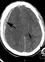
Axial multidetector computed tomography (MDCT) scan of the brain from a motor vehicle accident victim who died from multisystem blunt trauma. This axial MDCT scan shows a hyperattenuating acute right subdural hematoma (arrow). There is diffuse edema of the right cerebral hemisphere, compression of the ventricular system, and subfalcine herniation.
Acute epidural and subdural hematomas are classically hyperattenuating on MDCT scans, but there may be mixed attenuation if multiple bleeding episodes occurred before death or if there is decomposition. Chronic subdural hematomas typically demonstrate fluid attenuation on MDCT scans, because they are composed of serosanguineous fluid. Small subdural hematomas that are thinly layered beneath the dura may be difficult to appreciate.
Diffuse axonal injury is a difficult diagnosis to make on MDCT scanning. The brain often appears normal, but it may show petechial hemorrhages in the corpus callosum and at the gray-white junction. Subarachnoid hemorrhage is present in most cases of moderate to severe head trauma. It is seen as a thin layer of high attenuation in the cerebrospinal fluid (CSF) spaces, cisterns, and sulci on MDCT scans, as depicted in the following image.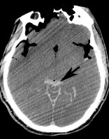
Axial multidetector computed tomography (MDCT) scan of the brain from a motor vehicle accident victim who died from multisystem blunt trauma. This axial MDCT scan shows subarachnoid hemorrhage adjacent to the cerebellar vermis (arrow). A small amount of intraventricular hemorrhage is also present. The intracranial gas and loss of gray and white matter differentiation is due to decomposition.
Decomposition makes the diagnosis of subarachnoid hemorrhage more challenging, because the dura adjacent to the brain appears relatively dense as decomposition begins to occur, and blood decreases in attenuation as it decomposes.
Pulmonary lacerations may appear as focal consolidations or cavities on MDCT scans. Linear tracks of gas through the lung may also indicate communication with a bronchus and an associated tracheal or bronchial laceration. Hemorrhage in the mediastinum is indicative of a major vascular injury. Radiography may show widening of the mediastinum from a periaortic hematoma, blurring of the aortic contour, or thickening of the paratracheal stripe.
On MDCT scans, aortic lacerations are characterized by alteration in the position and contour of the aorta. MDCT angiography is potentially useful to identify the site of rupture. Injuries to the aortic arch branches, pulmonary artery, and vena cava may also produce mediastinal hematomas. The location of hemorrhage may help establish the site of vascular injury. Diaphragm elevation should raise concern for diaphragm laceration or rupture. Intraabdominal organs may protrude into the thorax when there is laceration or rupture of the hemidiaphragm.
Diagnosis and interpretation of spine, pelvic, and extremity fractures is easily established with radiography and MDCT scanning. Fractures are linear, angulated, or displaced lucencies within bone, as demonstrated in the following 2 images.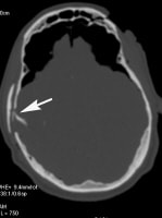
Axial multidetector computed tomography (MDCT) scan of the brain from a motor vehicle accident victim who died from multisystem blunt trauma. This axial MDCT scan shows a depressed skull fracture of the right parietal temporal region (arrow) and fracture of the left frontal sinus with overlying soft-tissue defect. The brain has retracted from decomposition, and there is decompositional gas within the cranium.
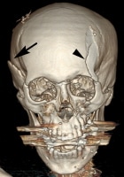
Three-dimensional multidetector computed tomography (MDCT) scan of the head from a motor vehicle accident victim who died from multisystem blunt trauma. This MDCT scan shows the right-sided depressed skull fracture (arrow) and a left frontal fracture that extends to the orbit (arrowhead). There is streak artifact from dental restoration.
The margins of acute fractures are well defined and lack sclerosis. In the skull, they may cross sutures and vascular impressions. Vertebral body compression fractures are characterized by loss of vertebral body height and/or increased density within the bone from the compressive forces. Vertebral body compression fractures and abnormalities in alignment are best viewed on sagittal MDCT images. Axial images are useful to view the pedicles and posterior elements of the vertebral bodies. Three-dimensional images provide an excellent depiction of the anatomic distribution of spine and pelvic fractures, which can be difficult to appreciate at autopsy.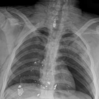
Frontal radiograph of the chest from a victim of a gunshot wound to the chest. This image shows metallic bullet fragments overlying the heart and right lower chest. There are right posterior rib fractures and a bilateral pneumothoraces.
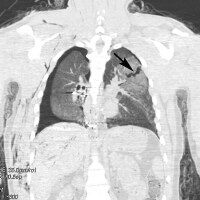
Coronal multidetector computed tomography (MDCT) scan of the chest from a victim of a gunshot wound to the chest. This image shows a gas-filled gunshot wound track that extends from the left upper lobe (arrow) to the right lower lobe. Metallic bullet fragments are located in the right lower lung and adjacent to the right hemidiaphragm. The increased density surrounding the gunshot wound track is hemorrhage.
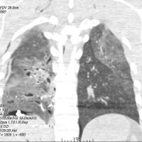
Coronal multidetector computed tomography (MDCT) scan of the chest from a victim of a gunshot wound to the chest. This image shows a gas-filled gunshot wound track that extends from the left upper lobe to the right lower lobe. Metallic bullet fragments are located in the right lower lung and adjacent to the right hemidiaphragm. The increased density surrounding the gunshot wound track is hemorrhage.
If the bullet passes through bone, bone fragments may also be present along the track. In the lungs, the finding of hemorrhage and cystic spaces characterize the bullet path. Gunshot wounds through the brain are characterized by hemorrhage, foci of gas, metallic fragments, and bone. In some cases, a distinct linear track within the brain is not identifiable. Metallic fragment analysis and the pattern of fragment deposition along the gunshot wound track are excellently depicted on 2-dimensional multiplanar and 3-dimensional images that have thresholds adjusted for metal attenuation. The various components of a bullet can be differentiated on CT scans, which facilitates recovery of the fragments for ballistic analysis.
The evaluation of skin-surface characteristics with MDCT scanning is limited. Three-dimensional MDCT algorithms can depict the entry and exit wounds, but skin-surface features such as wound shape, pigmentation, discoloration, and soot deposition are findings that can only be made on external examination of the body. (Gunshot wounds will be discussed in more detail in a separate article.)
Diffuse subarachnoid hemorrhage is characterized by high attenuation throughout the subarachnoid spaces that interdigitate between the cerebral gyri and in the basilar cisterns. Cerebral aneurysms are the most common cause of subarachnoid hemorrhage. The predominant location of subarachnoid hemorrhage on MDCT scans may be a clue to the location of the aneurysm, because the aneurysm may not be identified directly on routine postmortem MDCT images. Postmortem angiography has the potential of demonstrating the aneurysm and site of rupture.
If epilepsy is the cause of sudden death, the brain is often normal on MDCT scans. The role of postmortem imaging in these deaths is to exclude intracranial pathology that may be a seizure focus such as occult trauma, hemorrhage, or tumor, and to exclude other causes of death. Tumors and cerebral infarctions are most often viewed as low attenuation on noncontrast postmortem MDCT images. They may exhibit mass effect from vasogenic edema, and evidence of cerebral herniation may be present.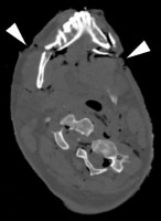
Axial multidetector computed tomography (MDCT) scan from an aviation accident victim who died from blunt trauma before the fire of the crash. This image of the lower face and neck shows a complex cervical spine fracture dislocation with transection of the cervical cord. Mandibular fractures are also present. Note the presence of full-thickness burns by the irregular contour of the subcutaneous fat and focal areas of thermal tissue loss (arrowheads).
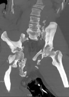
Coronal multidetector computed tomography (MDCT) scan from an aviation accident victim who died from blunt trauma before the fire of the crash. This maximum intensity projection image shows a complex fracture of the sacrum, pelvis, and right femur.
Full-thickness burns show loss of the dermal layer with exposure of the underlying fat and/or skeletal muscle that is typically irregular and jagged. In severely charred victims, skeletal muscle is exposed and retracted from shortening and thermal destruction of muscle, as seen in the image below.12 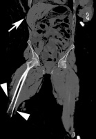
Coronal multidetector computed tomography (MDCT) scan of full-thickness burns and thermal amputation in a motor vehicle crash victim. This image of the abdomen and lower extremities shows thermal tissue loss with full-thickness burns of the abdominal wall (arrow) and lower chest. There is extensive thermal tissue loss of the lower extremities and thermal amputation of the right distal femur. Note the mottled lucency of the marrow space, as well as skeletal muscle retraction with exposed distal bone that is characteristic of thermal injury (arrowheads).
Thermal flexion deformities may occur in severely charred bodies, and these are associated with fractures and dislocations from the mechanical forces associated with muscular contraction and shrinkage. Thermal fractures are fine, linear cortical fractures in bone uncovered by soft tissue or bone that are typically found in areas of severe charring. In contrast, traumatic fractures are found in unexposed bone and are typical of mechanical injury, such as spinal compression fractures and pelvic bone fractures.
Although traumatic long bone fractures may be difficult to differentiate from thermal fractures, fractures in areas without charring and angulated fractures suggest a traumatic origin. The combination of retraction, flexion, dislocation, and fracture should facilitate the recognition that the findings are due to thermal injury rather than injury that occurred before death or before the fire.12
The visibility of a wound on MDCT scans is dependent on the orientation and position of the wound on the body. Skin wounds are characterized by a break in the continuity of the skin and are outlined by air. The track in the body is visible if air is carried into soft tissue or released from a gas-containing organ such as lung or bowel (see the image below)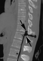
Sagittal multidetector computed tomography (MDCT) scan of a stab wound to the back that penetrates the spinal column. This image shows air in the wound track. The wound has a horizontal orientation and passes between the posterior vertebral elements and into the spinal canal, severing the spinal cord. The ends of the cord are retracted (arrows). There is no break in the skin surface, which is likely due to closure of the wound from the supine positioning of the body in the MDCT scanner.
By viewing sequential images on a workstation or using multiplanar reconstructions it may possible to determine the orientation and approximate length of the wound at the skin surface. It may also be possible to estimate the depth of the wound if the wound track contains gas or if there is adjacent bone injury. Air from venous air embolism may be seen in the right heart and venous structures following stab wounds to the neck or wounding of any major vein that permits air to enter the venous system.14
Internal hemorrhage may be seen in anatomic spaces where blood accumulates in sufficient volume. Hemopneumothorax, hemoperitoneum, perinephric hematoma, and subcapsular hemorrhage in intraabdominal organs are readily identified on MDCT images. Injuries to the heart are likely to cause hemopericardium with cardiac tamponade.14 The depth and direction of the wound may be estimated by detecting injury to underlying bone and soft-tissue structures.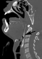
Sagittal multidetector computed tomography (MDCT) scan of a victim with sand aspiration in drowning. This image shows high-density sand throughout the pharynx.
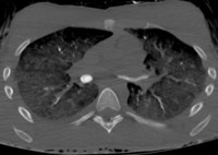
Sagittal multidetector computed tomography (MDCT) scan from a victim with sand aspiration in drowning. This image of the chest shows sand filling the right and left bronchi. Severe pulmonary edema is present.
The presence of sinus and airway fluid is a very nonspecific finding, because it may found in other forms of death or from decomposition. However, the presence of airway froth and sand may be helpful indicators of drowning. Airway froth is characterized by heterogeneous low attenuation fluid admixed with rounded foci of air. Sand, silt, or mud appears as high attenuation material within the sinuses or airways on MDCT images. Pulmonary edema is the most prominent lung finding of drowning on plain film radiography and MDCT scans. In mild pulmonary edema, there are interstitial and septal lines, as depicted below. With more severe edema, alveolar edema is present. Dilated and engorged right-sided cardiac chambers and great vessels may also be present. Even though these are nonspecific findings indicative of hypervolemia, they are helpful supportive findings when other anatomic findings of drowning are present.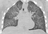
Coronal multidetector computed tomography (MDCT) scan from a victim with pulmonary edema in drowning. This image of the lungs shows moderate pulmonary edema evenly distributed throughout the lungs. Prominent septal lines are present in the bases and apices.
Watery fluid similar to that of the drowning medium may be found in the stomach at autopsy. Sand, silt, or other debris from the drowning fluid may be present as well. The amount of fluid is variable. Consequently, the degree of fluid distention of the stomach on MDCT images is not a reliable indicator that drowning fluid has been ingested at the time of death.Conclusions
Multimedia

Media file 1: Axial multidetector computed tomography (MDCT) scan of the brain from a motor vehicle accident victim who died from multisystem blunt trauma. This axial MDCT scan shows a hyperattenuating acute right subdural hematoma (arrow). There is diffuse edema of the right cerebral hemisphere, compression of the ventricular system, and subfalcine herniation. 
Media file 2: Axial multidetector computed tomography (MDCT) scan of the brain from a motor vehicle accident victim who died from multisystem blunt trauma. This axial MDCT scan shows subarachnoid hemorrhage adjacent to the cerebellar vermis (arrow). A small amount of intraventricular hemorrhage is also present. The intracranial gas and loss of gray and white matter differentiation is due to decomposition. 
Media file 3: Axial multidetector computed tomography (MDCT) scan of the brain from a motor vehicle accident victim who died from multisystem blunt trauma. This axial MDCT scan shows a depressed skull fracture of the right parietal temporal region (arrow) and fracture of the left frontal sinus with overlying soft-tissue defect. The brain has retracted from decomposition, and there is decompositional gas within the cranium. 
Media file 4: Three-dimensional multidetector computed tomography (MDCT) scan of the head from a motor vehicle accident victim who died from multisystem blunt trauma. This MDCT scan shows the right-sided depressed skull fracture (arrow) and a left frontal fracture that extends to the orbit (arrowhead). There is streak artifact from dental restoration. 
Media file 5: Frontal radiograph of the chest from a victim of a gunshot wound to the chest. This image shows metallic bullet fragments overlying the heart and right lower chest. There are right posterior rib fractures and a bilateral pneumothoraces. 
Media file 6: Coronal multidetector computed tomography (MDCT) scan of the chest from a victim of a gunshot wound to the chest. This image shows a gas-filled gunshot wound track that extends from the left upper lobe (arrow) to the right lower lobe. Metallic bullet fragments are located in the right lower lung and adjacent to the right hemidiaphragm. The increased density surrounding the gunshot wound track is hemorrhage. 
Media file 7: Coronal multidetector computed tomography (MDCT) scan of the chest from a victim of a gunshot wound to the chest. This image shows a gas-filled gunshot wound track that extends from the left upper lobe to the right lower lobe. Metallic bullet fragments are located in the right lower lung and adjacent to the right hemidiaphragm. The increased density surrounding the gunshot wound track is hemorrhage. 
Media file 8: Axial multidetector computed tomography (MDCT) scan from an aviation accident victim who died from blunt trauma before the fire of the crash. This image of the lower face and neck shows a complex cervical spine fracture dislocation with transection of the cervical cord. Mandibular fractures are also present. Note the presence of full-thickness burns by the irregular contour of the subcutaneous fat and focal areas of thermal tissue loss (arrowheads). 
Media file 9: Coronal multidetector computed tomography (MDCT) scan from an aviation accident victim who died from blunt trauma before the fire of the crash. This maximum intensity projection image shows a complex fracture of the sacrum, pelvis, and right femur. 
Media file 10: Coronal multidetector computed tomography (MDCT) scan of full-thickness burns and thermal amputation in a motor vehicle crash victim. This image of the abdomen and lower extremities shows thermal tissue loss with full-thickness burns of the abdominal wall (arrow) and lower chest. There is extensive thermal tissue loss of the lower extremities and thermal amputation of the right distal femur. Note the mottled lucency of the marrow space, as well as skeletal muscle retraction with exposed distal bone that is characteristic of thermal injury (arrowheads). 
Media file 11: Sagittal multidetector computed tomography (MDCT) scan of a stab wound to the back that penetrates the spinal column. This image shows air in the wound track. The wound has a horizontal orientation and passes between the posterior vertebral elements and into the spinal canal, severing the spinal cord. The ends of the cord are retracted (arrows). There is no break in the skin surface, which is likely due to closure of the wound from the supine positioning of the body in the MDCT scanner. 
Media file 12: Sagittal multidetector computed tomography (MDCT) scan of a victim with sand aspiration in drowning. This image shows high-density sand throughout the pharynx. 
Media file 13: Sagittal multidetector computed tomography (MDCT) scan from a victim with sand aspiration in drowning. This image of the chest shows sand filling the right and left bronchi. Severe pulmonary edema is present.
Sunday, October 17, 2010
(Enlarge Image)
(Enlarge Image)
(Enlarge Image)
(Enlarge Image)
(Enlarge Image)
(Enlarge Image)
(Enlarge Image)
(Enlarge Image)
(Enlarge Image)
(Enlarge Image)
(Enlarge Image)
(Enlarge Image)
(Enlarge Image)
Labels: Postmortem Radiology and Imaging
Subscribe to:
Post Comments (Atom)
0 comments:
Post a Comment